On November 14th the Alberta section of the Microscopy Society of Canada was fortunate to host a presentation by Dr. Yuji Konyuba from JEOL Ltd. (Akishima, Japan). See Fig. 1. Dr. Konyuba presented a talk entitled “Progress in Cameras for Transmission Electron Microscopy”. Dr. Konyuba has provided a deep background of the physics principles, as well as technology and engineering challenges encountered in design and manufacturing of modern cameras for transmission electron microscopy. He summarized and compared practical advantages and drawbacks of the most frequently used set ups, such as the signal collection efficiency, sources of artifacts, frame rate and cost. He has discussed side mounted cameras, that can a particularly cost-effective solution, and on bottom mount cameras with scintillator-fiber optics stack, see Fig 2. The direct exposure, back-thinned chips were discussed only briefly.
In the later part of his talk, Dr. Konyuba dived in discussion of a camera he developed at JEOL, the SightSKY. The camera has number of features that make it suitable for both materials sciences and biological transmission electron microscopy. In particular, the high frame rate, 58 frames per second (fps), at full 19 megapixel resolution. The high number of pixels allows to collect large field of view data while collecting and simultaneously observing in real time high pixel resolution data, Fig 2a.
The 58 fps frame rate is used always to read the camera, but to display the data, the frame rate can be adjusted by for example aligning and summing subsequent frames. The rather high frame rate and high detection quantum efficiency make the camera suitable for observation of radiation sensitive materials, although competing direct electron detection technology is likely to provide additional benefit at an incremental cost. The high frame rate is also of use to reduce the effect of sample drift. In development of new cameras of the scintillator-fiber optics stack type, a significant effort goes to optimizing the scintillator properties, such as the amount of light per incident electron, the point spread function and the avoidance of artifacts arising from the structure of the fibre optics stack. When the entire system is optimized, both materials science and a rather low dose observations of biological samples can be performed, Fig. 2b).
About 45 people participated, both in person and over zoom. This is a high number considering that Remembrance long weekend ended on November 13, 2023.
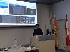
Figure 1. a) Dr. Yuji Konyuba presenting a talk on development and applications of cameras for transmission electron microscopy. On the displayed slide one can see example results of the SightSKY demonstrating its performance in electron diffraction and imaging modes. The high number of pixels allows to observe large field of view while acquiring data that can be inspected for detail at high pixel magnification simultaneously and in real time.
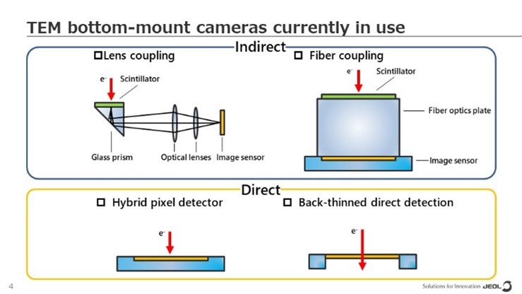
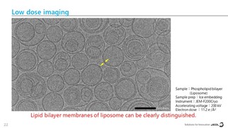
Fig 2. a) Schematic depiction of types of cameras. Top panel are coupled camera, either side mounted with a mirror, or coupled using a fiber optics stack. b) Low dose image acquired using the SightSKY.