H. Xu* , B. C. Choi* , Th. Speliotis† and Y. K. Hong‡
*Department of Physics and Astronomy, University of Victoria, Victoria, BC, V8W 3P6, Canada
† Institute of Materials Science, NCSR “Demokritos” 15310 Aghia Paraskevi, Greece
‡ Department of Electrical and Computer Engineering, and MINT Center, University of Alabama, Tuscaloosa, AL 35487, USA
Magnetic thin films have been the subject of extensive research owing to their applications in magnetoelectronic devices such as hard drive read heads [1] and magnetic random access memories [2]. Increasing demands in speed and storage capacity of magnetoelectronic devices makes it necessary to have a thorough understanding of magnetic media at sub-micron/sub-nanosecond scales.
Both quasi-static and transient magnetic behaviors of lithographically patterned, 10 μm-sized ferromagnetic squares were studied using Kerr effect microscopy. As a polarized laser beam reflects off a magnetized surface, its plane of polarization rotates by an angle θ proportional to the surface magnetization (Kerr rotation). θ , and hence the local magnetic configuration can be measured using photodiodes after passing the laser beam through a polarizer-analyzer pair oriented at extinction. Time-resolved measurements were performed using a stroboscopic pump-probe setup [3], with an pulsed Ti:sapphire laser serving as the magneto-optic probe, and a nanosecond magnetic pump field triggered synchronously by the laser with an electronically-controlled delay, at temporal resolutions of ~50 ps. Magnetic domain images were recorded by raster scanning the sample surface with a computer-controlled piezo flexure stage, at a spatial resolution of ~1 μm (Rayleigh diffraction limit).
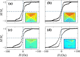
Figure 1 shows the measured quasi-static magnetic hysteresis for four different samples. The difference in their hysteretic behavior can be explained by the corresponding difference in their remnant magnetic configuration (inset), imaged using a scanning Kerr effect microscope.
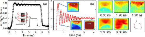
Figure 2 shows examples of spatiotemporal magnetic signals in response to a nanosecond magnetic excitation field, measured with the time-resolved scanning Kerr microscope. Average magnetization undergoes large angle switching towards the direction of applied field, followed by damped gigahertz oscillations (2(a)). Insight into the physical origins of observed magnetization dynamics can be obtained from non-equilibrium magnetic domain configurations, captured at different points during the excitation process. In 2(b), the alternating contrasts between the top and bottom quadrants in the domain images indicate out-of-phase behavior, which eventually damps out. 2(c) shows the gyrotropic motion of the magnetic vortex core (out-of-plane magnetization at the center of the sample) during the excitation.
[1] S. Parkin et al., Proceedings of the IEEE, 91(5) (2003) 661–680.
[2] J. G. Zhu et al., Journal of Applied Physics, 87(9) (2000) 6668. [3] M.R. Freeman et al., IEEE Transactions on Magnetics, 6 (1991) 4840.