Marek Malac and Misa Hayashida
The Alberta section hosted two excellent speakers last summer: Dr. Toshie Yaguchi from Hitachi High Technologies (Hitachinaka, Japan) on Aug 3rd , 2018 and Dr. Yuji Konyuba (JEOL Ltd. Akishima, Japan) on Aug 14th, 2018.
Dr. Yaguchi presented new results obtained from the latest 20 to 120 kV Hitachi analytical transmission electron microscope (TEM). The instrument builds on the success of the 7700 and 7800 series 120 kV instruments that were originally designed for biological applications. The new HT-7800 series instrument has several important features that make it suitable for materials science applications. In particular, the new objective lens with less than 0.2 nm resolution enables imaging of the c rystal lattice of many materials. The instrument can be operated between 20 to 120 kV, a feature that is very useful for imaging radiation sensitive materials and for contrast tuning, especially for thin and light element samples. In some materials, decrea sing the incident electron beam energy below a threshold can result in virtually damage - free observation of the sample. Since the instrument is based on a 120 kV column, it has a very small footprint and is more affordable than 200 kV or 300 kV instruments . Both parallel beam TEM and scanning TEM (STEM) modes are available in the 120 kV instrument. The LaB 6 electron source appears to be a good compromise. It is convenient to maintain and replace, and provides high current in parallel beam TEM mode, yet allo ws for reasonable resolution and acceptable beam current in the scanning TEM mode. To further increase the utility of the instrument , it is equipped for in-situ environmental TEM (ETEM) observations. The main changes include high speed turbo - molecular pump (300 l/s) combined with an oil free scroll pump and newly designed automatic gun air lock valve for environmental TEM allowing for in-situ gas-solid reaction observations.
The applications shown by Dr. Yaguchi included in-situ observation of the interac tions of electrocatalysts with their support. A small (1 μm diameter) selected area aperture allows for collection of parallel beam diffraction from sub - 20 nm area of the sample at low irradiation dose. In fact, the irradiation dose is several orders of ma gnitude lower than the dose needed for imaging, while the sub - 20 nm probed diameter is sufficient to study the structure and orientation of individual nanoparticles or a small region of the sample. Indeed, electron diffraction provides more reliable means for identification of sample structure than high resolution imaging, especially when coupled with real time software for diffraction pattern analysis, as implemented in 120 kV Hitachi instruments.
In-situ heating, up to 1500°C, is possible in the instrum ent at gas pressures up to ~0.3 Pa, using a specimen heating holder with a gas injection nozzle. A practical example included the study of fuel cell deterioration at elevated pressure in humid air environment . The most impressive example of the capabilitie s of this tiny, but powerful, instrument was in-operando imaging of a polymer electrolyte fuel cell. A newly developed holder enables measuring the voltage across the fuel cell while injecting H2 and O2 simultaneously and continuously on the anode and cath ode of the fuel cell, respectively. The observations of the fuel cell operation are then performed in real time. The image below shows the evolution of the cathode catalyst of the membrane electrode assembly [1]. In order to avoid any damage to the observe d structures, the electron beam irradiation condition was optimized and the images were taken at a low dose. However, even low dose TEM images provide useful information on the microstructure evolution. Most importantly, the result demonstrates the possibility of the simultaneous in-situ electrochemical measurement along with TEM observations.
[1] Takeo Kamino Toshie Yaguchi and Takahiro Shimizu, Development and Application of a Sample Holder for In Situ Gaseous TEM Studies of Membrane Electrode Assemblies for Polymer Electrolyte Fuel Cells , Microsc. Microanal. 23 (2017), p.945 – 950.
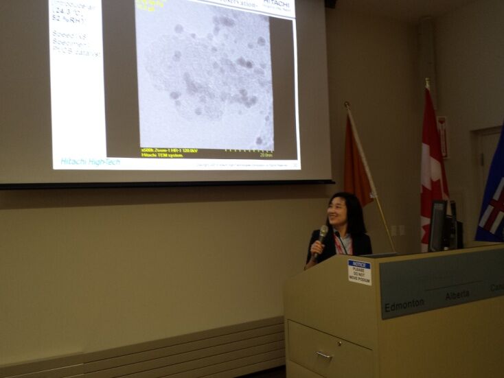
Dr. Yaguchi presenting for MSC Alberta section in Taylor room of NRC - NANO.
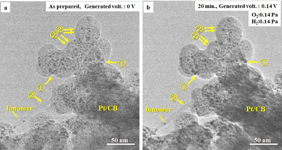
Evolution of polymer membrane fuel cell in-operando. The experiment was performed in Hitachi's newly developed 120 kV environmental TEM/scanning TEM. The as-prepared sample images in a) were collected under vacuum while b) was collected at 0.14 Pa of H2 and 0.14 Pa of O2 . The presence of additional scattering from gas in b) is visible in the vacuum area in the top left part of a). Following in-situ exposure to O2 and H2 the Pt nano particles on carbon black (CB) were partially agglomerated, marked with 1 in b). Furthermore, the edges of the Ionomer layer and carbon black support became darker, while the inside was brighter, marked with 2 in b).
Dr. Konyuba’s talk covered two main topics: phase contrast imaging and imaging of large area samples in a transmission electron microscope. Phase contrast imaging in TEM is gaining popularity as it provides contrast enhancements on a wide variety of samples. The concept of phase imaging in TEM is similar to phase plate imaging in an optical microscope. A phase shift between unscattered and scattered wave is induced by placing a suitable hardware in the back focal plane of the (transmission electron) microscope objective lens. The phase shift between the scattered and unscattered wave alters the contrast transfer of the microscope resulting in an increased contrast i n the final image. Implementation of a uniform film placed in the back focal plane of an objective lens is very easy to use. The method was jointly developed by NRC-NANO and JEOL and is referred to as hole - free phase plate (HFPP) [1] as an alternative to Z ernike phase plate imaging. In Zernike phase plate imaging in TEM a film with a hole is fabricated. During operation, the hole of the Zernike phase plate is centered on the unscattered electron beam resulting in a phase shift of the scattered beams due to presence of the sharp edge of the hole in the Zernike phase plate film. The core concept of the HFPP is that the electron beam itself is used to locally modify the properties of a uniform film placed in the back focal plane of the imaging lens, thus elimin ating the need for precise alignment of the beam and the hardware of the phase plate. The lifetime of a HFPP is very long because virtually any place of the uniform film can be used to generate phase plate effect. Furthermore, the HFPP eliminates the fring ing contrast that plagues Zernike phase plate imaging in TEM leading t o clean looking images. The image below shows a comparison of standard bright field TEM in a) and Zernike phase plate image in b). When compared to bright field TEM in a) the HFPP images show significant increase in contrast while they do not show the spurious fringing seen in ZPP images in b). Further examples of HFPP application included cryo-electron tomography of biological samples, images of block co-polymer, and visualization of sem iconductor chips. The last application is particularly powerful as HFPP allows to observe and differentiate, in focus, the contrast from different materials, such as Si oxides, leading to localized strong contrast in images of semiconductor devices. Dr. K onyuba also presented HFPP application for imaging block copolymers where the use of HFPP identifies the location of two carboneous components of the block co - polymer at low irradiation dose.
The large area sample refers to samples up to 1 x 2 mm2 mounted on custom-developed 30 nm thick SiN film. A sample, such as slices from microtome, can be deposited onto the SiN film and observed in a fully automated TEM, such as JEOL 1400 [2]. The use of microfabricated SiN membrane eliminates the substrate wr inkles that are observed in formvar support grids while the background from the 30 nm film is comparable to that of standard 80 nm thick formvar support film. The examples of large area observations included TEM imaging of Drosophylia embryo a 680 x 250 μm2 area with sufficient sampling to observe the cell membrane.
The talks were interesting and motivating and we look forward to hosting more presentations and workshops at the Alberta section in the future.
[1] Marek Malac, Marco Beleggia, Masahiro Kawas aki, Peng Li and Ray F Egerton, Convenient contrast enhancement by a hole-free phase plate , Ultramiroscopy, 118 (2012), p. 77-89.
[2] Yuji Konyuba, et al., Fabrication and characterization of sample - supporting film made of silicon nitride for large-area observation in transmission electron microscopy, Journal of Electron Microscopy, (2018), in press.
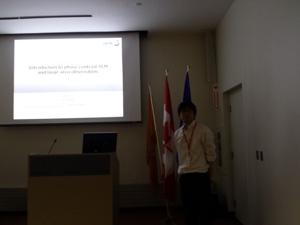
Dr. Konyuba presenting for MSC Alberta section in Taylor room of NRC-NANO.
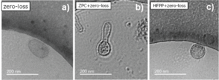
Imaging of exosome embedded in ice. a) standard bright field TEM image, b) Zernike phase plate image with increased contrast, but clearly visible fringing contrast artifacts, c) a hole-free phase plate (HFPP) image that exhibits significantly higher contrast relative to a) with no spurious fringing as visible in b). The images were collected at ~3 μm defocus and irradiation dose ~30 e-/Å2.